


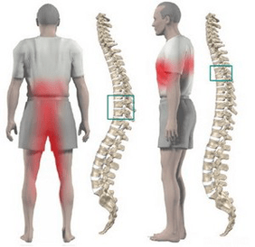
In the case of thoracic osteochondrosis, the organs associated with the areas of the spinal cord at and below the level of the affected thoracic region often suffer. Impairment of the normal functioning of the spine causes immobility of the arms, legs and torso as a whole, dysfunction of the pelvic organs, respiratory muscles and internal organs.
Osteochondrosis is a degenerative-dystrophic disease of the spine, which is based on the replacement of intervertebral discs with the involvement of the entire spinal apparatus in the pathological process of adjacent vertebrae and intervertebral joints.
Features of the anatomy of the spine
The mobility and stability, elasticity and resilience of the spine depend more on the intervertebral discs, which are one of the types of cartilaginous connections between bones and provide a strong connection between the bodies of adjacent vertebrae. The total length of the intervertebral discs is a quarter of the length of the spine.
The most important function of discs is to reduce the vertical load on the vertebrae. The disc consists of three parts:
- hyaline plates (closely adjacent to the vertebrae);
- nucleus pulposus (fills the space between the plates);
- fibrous ring (covers the core from the outside).
The nucleus contains cartilage cells, tightly bound collagen fibers, and chondrin (proteoglycans). The anterior surface of the discs is covered with an anterior longitudinal ligament that is tightly connected to the vertebrae and rotates freely on the discs. The posterior longitudinal ligament is firmly attached to the surface of the disc and forms the anterior wall of the spinal canal. The intervertebral disc does not have its own blood supply, so it is nourished by substances that diffuse from the spinal cord.
The distribution of vertical loads on the spine occurs due to the elastic properties of the discs. As a result of the pressure, the pulposus nucleus expands and the pressure is redistributed to the annulus fibrosus and hyaline plates. When moving, the nucleus moves in the opposite direction: when bent - to the convex, when not bent - to the front. As the spine moves, muscles, ligaments, and discs are involved. Therefore, a violation in one transition causes a violation in the entire kinetic chain.
Causes of the disease and the mechanism of its development
Mechanical impact on the spine plays a special role in the development of osteochondrosis. Under the influence of unfavorable static and dynamic loads, the pulposus nucleus gradually loses its elastic properties (as a result of depolymerization of polysaccharides), forming protrusions and sequestrators.
The process of disc degeneration is influenced by a genetic predisposition, which leads to the development of changes in the neuromuscular apparatus of the back, changes in the structure of glycosamines and disruption of the distribution of collagen fibers in the disc. In the development of thoracic osteochondrosis, a genetic factor that is subject to increased functional activity is very important.
Risk factors for the development of degenerative changes in the spine include the anatomical features of discs with evolutionary defects. One of these features is the nutritional properties of the structures. In the human body, the disc is composed of poorly perfused tissue. Closure of blood vessels occurs in infancy. Occurs due to the spread of substances from the last plates after feeding.
The stimulator of nutrient penetration is a dosed load that excludes static postures and great stress. Physical inactivity is one of the main risk factors for thoracic osteochondrosis. Therefore, regular exercise is an important preventive measure.
The peculiarity of the microscopic structure - a few cells - reduces the intensity of regenerative capacity and the rate of recovery of disk components. An anatomical feature is the weakness and lack of strength of the discs in the back. This contributes to the appearance of wedge-shaped discs in the lower thoracic and lumbar regions.
Involuntary changes in the development of osteochondrosis are of great importance. Active degenerative changes begin to increase after 30 years. The synthesis of components necessary for the disc (glycosaminoglycans) continues, but their quality deteriorates. Hydrophilicity decreases, fibrillation increases, sclerosis appears.
Stages of degeneration of intervertebral discs:
- long-term asymptomatic course, degenerative changes in intradiscal components, displacement of the nucleus inside the disc;
- open radicular symptoms of thoracic osteochondrosis, compression of the spinal cord, protrusion of the pulposus nucleus (protrusion, 1 degree);
- disc herniation with hernia protrusion (hernia, 2nd degree);
- degenerative changes in extradiscal components (grade 3).
Pathological protrusion compresses nerve roots, blood vessels or spinal cord at different levels (cervical, thoracic, lumbar) determines the clinical picture.
Restriction of thoracic lumbar mobility due to the presence of the chest contributes to minimal trauma to the intervertebral discs and therefore osteochondrosis. Physiological thoracic kyphosis helps to redistribute the weight of the upper half of the body to the lateral and anterior parts of the vertebrae. Therefore, intervertebral hernias and osteophytes form on the anterior and lateral surfaces of the spine. Posterior osteophytes and hernias are extremely rare.
Osteochondrosis helps to narrow the intervertebral foramina and compress the roots of the spinal cord and sympathetic fibers. Sympathetic fibers are formed in the gray matter of the spinal cord, then accumulate in the nodes and from there are sent to all internal organs. In addition to the typical neurological disorders of thoracic osteochondrosis, this leads to dysfunction of internal organs (vegetative, vasomotor, trophic) and imitation of somatic diseases. This feature of thoracic disc osteochondrosis explains the difficulties in diagnosing and prescribing the right treatment.
Symptoms of thoracic osteochondrosis
Thoracic osteochondrosis is more common in people who lead a sedentary lifestyle. At the same time, dosed loads do not have a stimulating effect on the spine, which contributes to the disruption of disc regeneration. Diseases are caused by prolonged work on the computer, bending, etc. such people should do therapeutic exercises independently.
Chest osteochondrosis often presents with dull pain, with less pain and a burning sensation. The pain is localized between the shoulder blades. The patient is worried about the feeling of chest tightness. When you feel the spinous processes of the thoracic vertebra, you find local pain that increases with axial loads on the spine, deep inspiration and rotations of the body.
Some patients have severe pain in the back and lower chest (posterior costal syndrome). This symptomatology develops as a result of displacement of the lower ribs. The pain increases sharply when turning the torso. More often, the pain syndrome disappears sharply.
Often, chest pain occurs in the girdle, corresponding to the course of the intercostal nerve. Sensitivity is impaired in the innervation zone of the corresponding nerve ending, paresthesias are visible, and a decrease in superficial and deep sensitivity is often observed. Possible impairment of abdominal press function, changes in knee and calcaneal tendon reflexes.
Internal organ dysfunction occurs when any nerve root is compressed from 1 to 12 at chest level. The thoracic region contains structures responsible for the innervation of the lungs, heart, intestines, liver, pancreas, and kidneys. Therefore, there are no symptoms that are unique to thoracic osteochondrosis.
The disease manifests itself with symptoms characteristic of another pathology:
- difficulty breathing;
- severe night pains;
- "heart", anginal pains;
- pain in the mammary glands;
- pain in the right or left hypochondrium (symptoms of cholecystitis and pancreatitis);
- pain in the throat and esophagus;
- pain in the epigastrium, abdomen (symptoms of gastritis, enteritis and colitis);
- sexual dysfunction.
Diagnostics
The greatest value in the diagnosis of thoracic osteochondrosis is a chest X-ray. The figure shows a decrease in the height of the intervertebral disc, sclerosis of the last plates, the formation of osteophytes.
Computed tomography allows to determine the condition of the vertebrae, the joints of the spine, the size of the spinal canal, to determine the location and size of the hernia.
When making a differential diagnosis, it is necessary to carefully collect a medical history and compare all the clinical signs of thoracic osteochondrosis with the symptoms of other diseases. For example, pain in the heart with osteochondrosis is not stopped by nitroglycerin, epigastric pain is not associated with food intake, it is not seasonal, all symptoms appear mainly in the evening and disappear completely after a night's rest.
How to treat thoracic osteochondrosis?
In almost all cases, treatment of thoracic lumbar osteochondrosis is conservative. The indication for therapy is the predominance of visceral syndromes with neurological disorders. The main orthopedic treatment should be adequate spinal traction:
- active vertical traction under water;
- Passive horizontal traction on the inclined bed using the Glisson curve by axillary straps during injury at the level of the thoracic vertebrae 1-4, 4-12 thoracic vertebrae.
Drug treatment consists of paravertebral blockades with novocaine solution. As the disease progresses, analgesics and sedatives are used. Ointments containing analgesics and anti-inflammatory drugs are allowed at home with unexplained pain syndrome.
After the elimination of acute events, massage of the muscles of the back and lower extremities is used. Manual therapy is indicated for 1-3 degrees of osteochondrosis in the development of functional blockages. There are a variety of options for soft and rough effects on the back muscles.
Therapeutic exercises allow you to dose all parts of the spine, which stimulates the recovery process. An important condition of exercise therapy for osteochondrosis is to exclude vertical loads.
Physiotherapy: UHF treatment, ultrasound, inductothermy, radon and pine-needle salt baths. Underwater traction and hydromassage are actively used in the spa stage.
Surgical treatment is rarely used. The indication for surgery is compression of the spinal cord with a piece of prolapse disc.